
Laboratory Microscopes


Manufacturer for Microscopes


Laboratory microscopes: Areas of application in the laboratory
Microscopes are mainly used in laboratories of science, research, development, industry and production. It is an essential laboratory instrument in biology, medicine, chemistry or material sciences. For example, our equipment is used in industrial, biological and medical laboratories for the examination of tissue and cell cultures, urine, blood, plasma, smears, microorganisms, bacteria, yeasts, fungi, mycoses or histological sections.
The areas of application for guided microscopes from Kern & Sohn, Helmut Hund, A. Krüss Optronic or Levenhuk include clinics, doctors' practices, research institutes, universities, technical colleges and schools. Our cost-effective and low-priced instruments are suitable for professional applications in cell and microbiology, veterinary and human medicine such as dermatology, urology, gynaecology or pathology.
Furthermore, our laboratory microscopes are used for the production of food and beverages in the food industry and breweries, as well as for the examination of waste water samples and sludge in sewage treatment plants. In industrial production, a microscope is also used for material examination and material testing.
Design and construction of a microscope
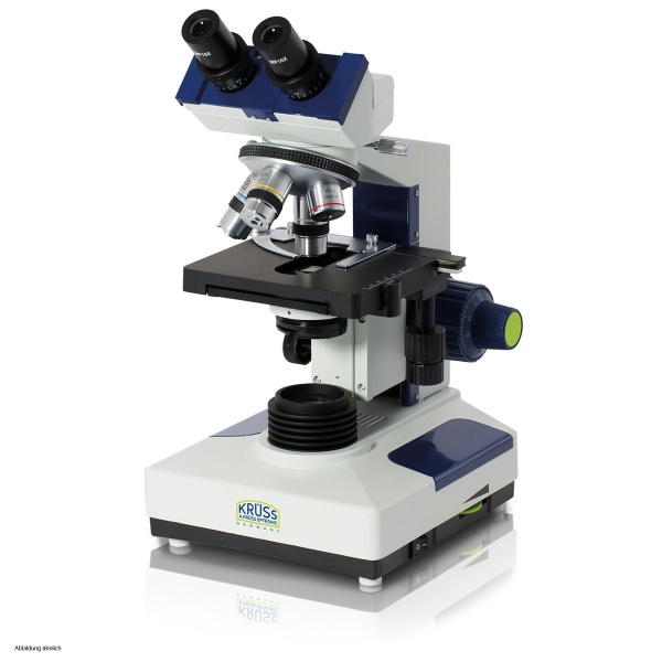
A microscope is an optical instrument for examining and magnifying very small objects. The instruments consist of two basic optical components, the objective and the eyepiece. These are connected to each other by the microscope tube.
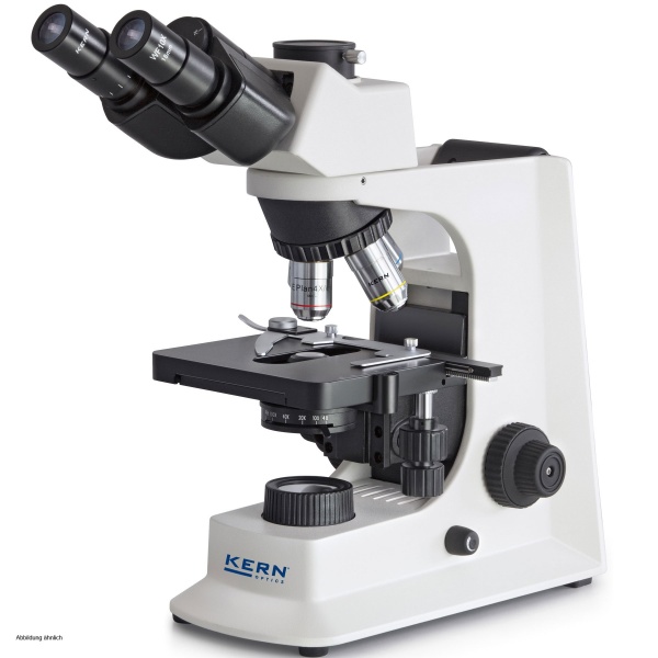
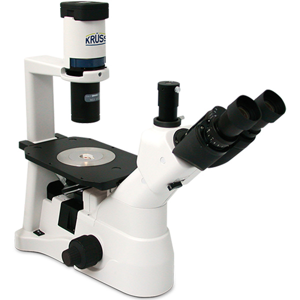
The various instruments differ depending on their design and field of application. In our online catalogue, we divide the laboratory microscopes into upright and inverted with regard to the construction.
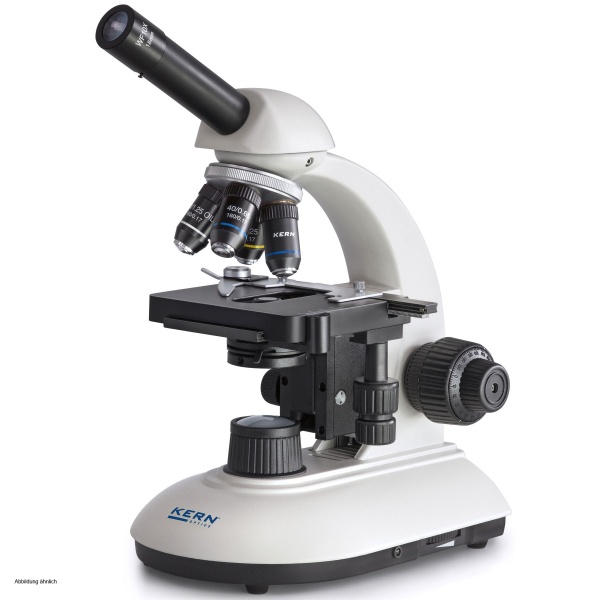
Essentially, a conventional microscope usually consists of the following components:
- Eyepiece - looking into, magnifies the intermediate image designed by the objective and projects it onto the retina of the eye;
- Tube - binocular, usually oblique tube at the top of the instrument;
- Objective - located on the instrument above the object being viewed;
- Revolving nose piece - allows different objectives to be changed quickly;
- Cross-stage - movable mechanical stage for exact positioning of the object to be viewed on the microscope stage;
- Specimen stage - centre-hole work surface including specimen clamps;
- Condenser - a system of lenses on the instrument below the specimen stage, optical light collector that concentrates the light;
- Condenser drive - drive knobs for focusing the illuminated and placed specimen;
- Illumination - usually a microscope light or mirror is used to illuminate the specimen;
- Illuminated field diaphragm - limits theilluminated area on the observed object of the microscopes;
- Stand with coarse and fine adjustment - usually two dials for focusing;
- Microscope base - base plate of the instrument.
ProfiLab24 assortment: Microscope types and their functions
In our online catalogue, we divide the microscopes offered by different manufacturers and models according to type, construction, objectives, illumination technology and constrating method.
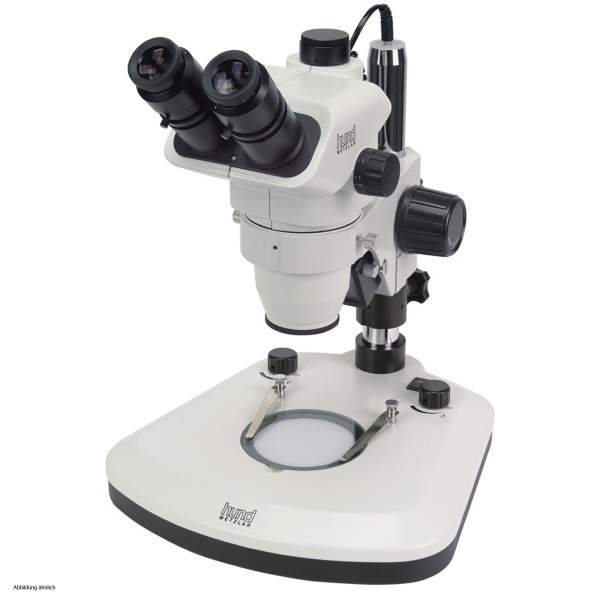
Microscope types
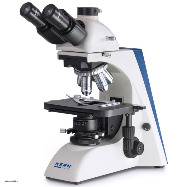
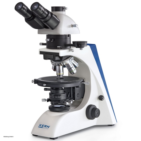
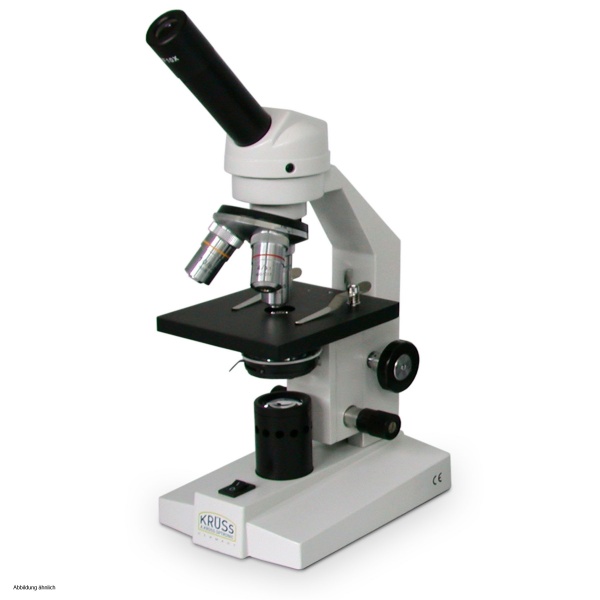
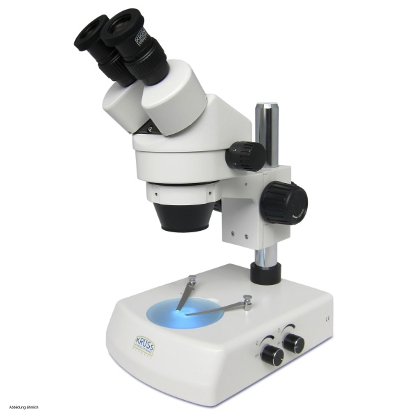
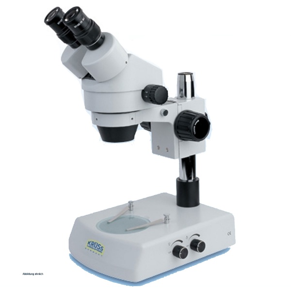
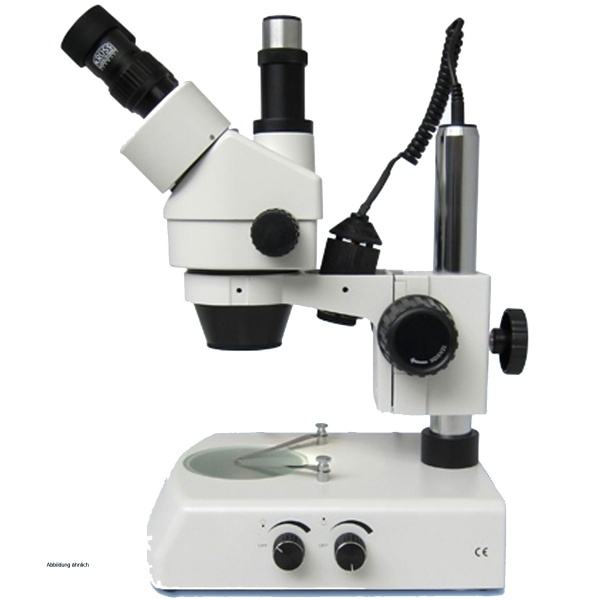
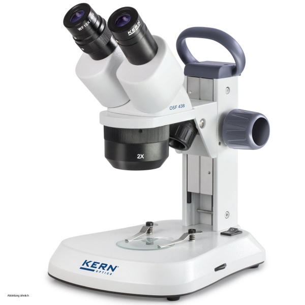
Currently, you will find stereo microscopes and monocular microscopes, binocular microscopes as well as trinocular microscopes in our portfolio. A stereomicroscope always has two eyepieces like any binocular microscope. With a stereomicroscope, the image is always designed simultaneously through two objectives that image the object from different angles, which is how the stereo effect is finally created. In a trinocular microscope, the third eyepiece is usually used with an additional vertically projecting phototube for attaching a camera.
Microscope objectives
Many characteristics of the microscopic image depend on the chosen objective of the microscope. We currently divide objectives into achromatic, epiplane, planachromatic, planachromatic-at-infinity-corrected and semi-planachromatic. Achromatic objectives, known as achromats, are usually the cheapest and most widely used objectives in microscopy due to their simple mechanical and optical design.
These devices usually consist of three lenses and are excellently suited for many routine microscopic examinations in the laboratory. The imaging errors of achromatic microscope objectives are not as extensively corrected as is the case, for example, with planachromatic microscope objectives, which eliminate the image field curvature that normally occurs in the microscopic image and produce higher contrast images.
Microscope illumination
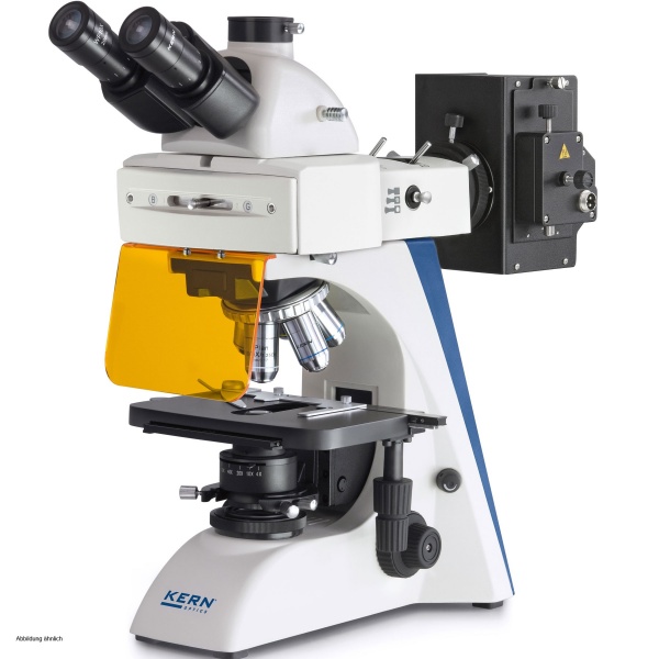
With regard to microscope illumination, we differentiate the instruments in our range into incident light, incident light fluorescence and transmitted light. In contrast to transmitted-light microscopes, reflected-light microscopes do not shine through the specimen being viewed. In reflected-light microscopes, the sample is illuminated from the direction of the objective - often through the objective itself.
Fluorescence microscopes use the physical effect of fluorescence for the microscopy of samples. In this process, fluorescent dyes are excited with light of one wavelength, causing light of another wavelength to be emitted a few nanoseconds later. Special filters ensure that only the emitted light is observed.
Microscopy Contrast method
With regard to the contrast method, we differentiate the microscopes offered into
- Incident light fluorescence,
- Incident-light brightfield,
- dark field,
- transmitted-light brightfield and phase contrast.
Many microscopic preparations, for example living cells or bacteria, are almost transparent in the unstained state and can hardly be seen with a classic brightfield microscope. This is why there are various contrast methods in microscopy, such as in front of a dark image background (dark field) or the widely used phase contrast method, for which special objectives and a condenser are required.
Accessories for your laboratory microscope
In our laboratory microscope assortment, we only carry high-quality microscopes from proven manufacturers. The robust microscopy solutions are characterised by their reliable quality, high diagnostic safety and service life as well as the functionality to exchange individual parts or to supplement them with accessory sets.
Depending on the model and manufacturer, you can order various spare parts and accessories for your microscope online, such as different objectives, attachments, filters, eyepieces, condensers, stands, specimen stages, specimen clamps, camera adapters, lighting elements as well as many other accessories and components for the guided microscopes.
Buy laboratory microscope online at ProfiLab24 - We are happy to advise you!
Do you still have questions or would you like individual advice on a laboratory microscope? No problem! We are always there for you! Simply contact us by phone at +49 30 403 667 940 or by e-mail at [email protected]. Together we will find the right microscope for your application.