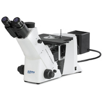
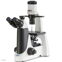
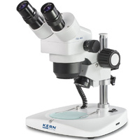
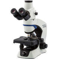
KERN OCM-1 inverted microscope
The KERN OCM-1 inverted microscope is a specialised inverted microscope for professional applications in cell biology, biotechnology and microorganism research. Thanks to its stable construction, flexible illumination and precise optics, it is particularly suitable for demanding routine work and research tasks. Compared to similar systems, it's especially handy when working with larger sample vessels and sensitive cell cultures, as a large working distance combined with ergonomic operation allows for the best results even during lengthy test series.
Precision and flexibility for demanding laboratory applications
The OCM-1 is primarily aimed at scientists and technicians in research laboratories, universities, pharmaceutical and biotechnology companies, and diagnostic facilities who regularly work with complex cell cultures and sensitive samples. The large working distance of 72 mm, in combination with the special Abbe condenser (numerical aperture 0.3), offers maximum flexibility for a wide variety of vessel shapes, such as culture flasks, Petri dishes or microtitre plates, while ensuring excellent image quality. The modular design allows for a wide range of adaptations to different methods - from classic bright-field microscopy to fluorescence analysis.
Technical highlights:
- Infinity optical system for consistently high image sharpness
- Large working distance (72 mm) with special Abbe condenser, N.A. 0.3
- Ergonomic, trinocular Siedentopf tube (45° inclination) with dioptre correction on both sides and variable eye relief (48-76 mm)
- 5-position objective turret for quick magnification changes (10x, 20x, 40x)
- Bright, continuously adjustable 30 W halogen transmitted light; OCM 165 with optional 100 W fluorescence illumination
- Mechanical object stage (Ø 118 mm) incl. holders for various cell culture vessels
- Robust construction, dimensions up to 782 x 304 x 530 mm depending on variant, weight from 14 kg
Further important details:
The inverse design principle ensures that objectives are positioned below the sample carriers. This allows even taller culture vessels to be analysed conveniently and directly - crucial for cell proliferation studies and long-term observations of living samples. The generous field of view (field number HWF, 2.2 cm) provides a broad overview when viewing specimens, which is particularly helpful for screening tasks or selecting particularly interesting cell colonies. Thanks to the modular design, a wide variety of lighting options (including HBO/LED reflected light or phase contrast unit) as well as various other eyepieces and objectives can be easily retrofitted and adjusted.
The mechanical stage is specially designed for quick changes between a wide variety of samples. The high stability of the microscope ensures low-vibration working conditions and precision, even during repeated, sensitive measurement series.
Certifications and standards
The microscope meets the strict requirements of professional laboratory environments and complies with the relevant CE, DIN and ISO standards for medical and biological laboratory equipment. This ensures safe and reliable operation in accredited facilities.
Specific application examples
-
Cell culture monitoring: Observation of mammalian cells during growth, evaluation of confluence or morphological changes in culture flasks or Petri dishes.
-
Microbiological screening: Analysis of bacterial and yeast cultures in microtitre plates, for example in antibiotic trials or drug screening.
-
Fluorescence-based methods: Detection of cell nucleus staining (e.g. DAPI), detection of specific antibody markers (immunofluorescence) or FISH analyses for gene localisation.
-
Tissue diagnostics and research: Documentation and evaluation of histological preparations, for example in the context of tumour research or diagnostics in pathology laboratories.
-
Toxicity studies and pharmacology: Real-time examination of cellular reactions to active substances or nanoparticles.
In the fluorescence range, dyes such as DAPI, FITC or TRITC are used to make cellular or subcellular structures visible.
FAQs
Difference between inverted and upright microscopes
In inverted microscopes, the objectives are located below the sample and the illumination units above it - ideal for observing cells in tall culture flasks/petri dishes.
Is fluorescence equipment absolutely necessary?
No, the fluorescence unit is only an additional option for special applications (e.g. immunofluorescence, FISH). Standard halogen illumination is sufficient for routine observations.
Which sample and vessel formats can be used?
The variably designed stage securely holds dishes, flasks and microtitre plates of various formats. Holders for different vessels enable quick adjustment.
How is the illumination intensity adjusted?
The brightness of the 30 W halogen transmitted light illumination is continuously adjustable - ideal for samples with different sensitivities and varying magnifications.
Ergonomics - suitable for longer working periods?
The 45° inclined trinocular Siedentopf tube prevents tension. Individually adjustable eye relief and dioptre correction support comfortable working even during long observation sessions.
Can the optics be individually expanded?
Yes, various objectives, eyepieces and other accessory options are available to flexibly adapt the system to specific laboratory requirements.
Conclusion
This inverted microscope sets standards for research and routine applications in cell biology, biotechnology and medical diagnostics: High-quality infinity optics, flexible illumination options and a sophisticated, ergonomic design support precise and efficient workflows. A robust, maintenance-friendly construction, coupled with a wide range of expansion options, makes the system an outstanding and long-lasting investment for any modern laboratory environment.